Latest Dental Technology
State of the art dental practice in Carlisle
We have four fully equipped, state of the art treatment rooms which all have temperature control facilities. The latest digital X-ray imaging equipment is available in each surgery. This investment enables our clinicians to deliver high quality treatment in a safe environment.
Operating microscope
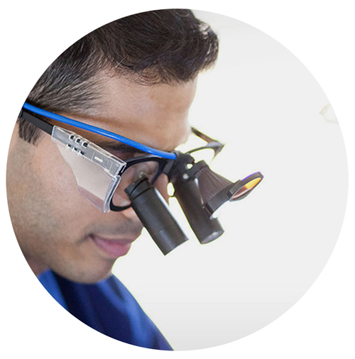
A dental microscope is unparalleled in its ability to provide an intensely illuminated, detailed and magnified image of the operating site. Campbell & Wilson Dental Practice is now equipped with a Surgical Operating Microscope to assist in performing the technical aspects of root canal treatment in particular. The most difficult aspect of root treatment is usually when accessing and identifying all the root canals. High-level magnification is absolutely essential in performing the difficult task of removing obstructions in the root canal.
The microscope can also be used to identify hairline cracks in teeth and aid our understanding of the complex root apex anatomy.
Digital radiographs
Digital radiographs enable delivery of treatment to be of the highest standard and involves the patient from the start. It is a form of X-ray imaging, where digital X-ray sensors are used instead of traditional photographic film. Using this system, the electronic sensor produces computerised radiographs, which appear instantly on the computer screen. The advantage of digital radiographs is that these images can be stored electronically, printed and utilised to communicate with other practitioners as required. The X-ray radiation dose is reduced by up to 80% compared to conventional film X-rays.
Cone Beam CT scans
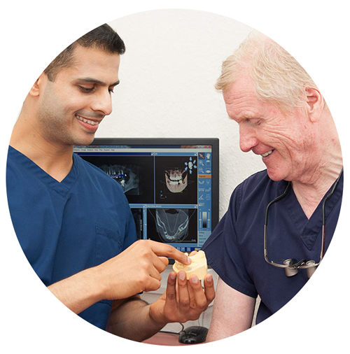
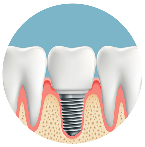
Dental radiographs only provide two-dimensional images but with CT scans we are able to view the structures of the mouth in three-dimensions. This is particularly useful for implant treatments to visualise the anatomy of the nerves, roots, bone and sinus. The CT scan is used to accurately determine the volume of bone available for placement of dental implants and helps us plan safe and predictable treatment.
We have a cone beam CT scanner at our practice in Penrith. The scan takes a couple of minutes and is entirely painless.
Computer guided Implant surgery
Computer guided implant surgery involves using computer technology to fit dental implants. Specialist software is required to visualize and virtually plan the implant placement procedure. A surgical guide can then be 3D printed and used on the day of surgery to ensure a more accurate implant placement. It increases the accuracy of the procedure as the surgeon can predetermine the best possible site for implant placement. It can simplify treatment and reduce complications.


